
Creating a functional vascular network in-vitro (VesselNet) based on anastomosis between large blood vessels and small capillaries:
We have demonstrated the role of a functional vascular network in-vitro in enhancing function and integration of implanted engineered tissues (Koffler et. al, PNAS, 2011 , Ben Shaul et al, PNAS, 2019). We showed that engineered vascularized muscle tissue cultured for 3 weeks before implantation, matured and integrated much better than grafts cultured for just 1 week before implantation (A-B).
In vitro culture time and vessel maturity levels positively correlated with integration dynamics of vascularized graft; constructs with more mature vessels integrated faster and the vasculature became more functional, with fewer clotting events in the graft (C-E).
Based on our previous experience in engineering vascularized tissues using flap techniques Shandalov et al, PNAS, 2014 , Freiman et al, J. Tissue Eng.Regen.Med, 2017, Egozi et al, J.Vis.Exp. 2016) we believe that the engineered in-vitro functional network should contain interconnecting large and small vessels.
Significant vascularization of pre-vascularized constructs containing small capillaries occurs when implanted wrapped around large vessels (artery and vein). Sprouts from these large vessels anastomosed with the small capillaries of the construct, resulting in a well vascularized construct (F). We intend to mimic this vascularization process in an in-vitro settings.
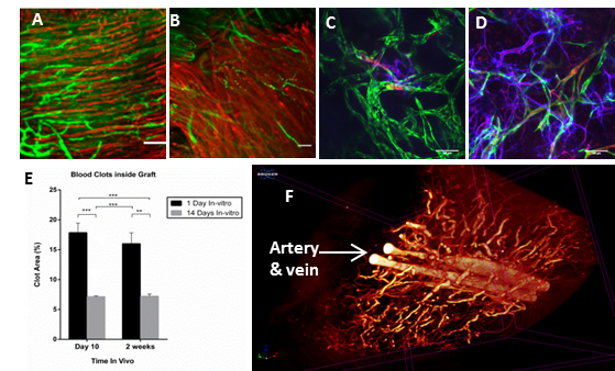
Supporting evidence for creating functional vasculature in-vitro. (A-B) Muscle fiber organization (desmin staining – red) , and integration with host vessels, marked by dextran perfusion (green), of implanted construct cultured for 3 weeks in-vitro before implantation (A) compared to construct cultured for 1 week (B). Constructs cultured for 3 weeks before implantation resemble native muscle organization. (C-D) Maturation and host integration of a 14-day-in-vitro cultured vascularized graft (D) compared to 1-day-cultured graft (C), host vessel – blue, engineered vessel – green, dextran-red. (E) Graft clotting as observed 10 and 14 days post-implantation in grafts cultured for 1 versus 14 days before implantation. (F) Micro-CT image presenting in-vivo vascularization of a construct vascularized with small capillaries wrapped around an artery and vein. Sprouts from artery and vein anastomosed with the small capillaries.